Home
/ Bone Cross Section Histology - Bone Wikipedia : There are two ways to study bone histology.
Bone Cross Section Histology - Bone Wikipedia : There are two ways to study bone histology.
Bone Cross Section Histology - Bone Wikipedia : There are two ways to study bone histology.. Bone is hard and many of its functions depend on that characteristic hardness. Most features of bone (but not the canaliculi, Spongy bone, also known as cancellous bone or trabecular bone, looks like a sponge under the microscope. Image.shutterstock.com if you look at the cross section of a long bone under a microscope, the rings of bone immediately internal to the periosteum of the bone are called _____. At the outer regions of the section, you can see a dense, thick layer of compact bone.
Histology sauropod vertebra picture of the week these pictures of this page are about:long bone cross section. This photo shows a cross section through bone. Spongy bone, also known as cancellous bone or trabecular bone, looks like a sponge under the microscope. Dry bone is cut and polished before mounting on a slide. Muscle attachments are visible along the outer surface.
Histology Review Bone Tissue Dr Tim Ballard Department Of Biology And Marine Biology Ppt Download from images.slideplayer.com Spongy bone also contains osteocytes housed in lacunae, but they are not arranged in concentric circles. Spongy bone is the osseous tissue, which fills the interior cavity of bones, consisting of mineralized bars called there are two ways to study bone histology. In addition to discussing the cellular constituents of bone and the architectural arrangement of their products, this article will also address the embryology and mechanisms of ossification as well. Related to bone cross section histology. Dry bone is cut and polished before mounting on a slide. Fetal leg, cross section, h&e, 40x (bone marrow in tibia and fibula, developing blood cells, sinusoid 37829 x 41067, megakaryocyte 37861 x 39647, 38143 x 39087, 39555 x 36969, 31707 x 18214). This section will examine the gross anatomy of bone first and then move on to its histology. This is a cross section through decalcified bone.
Figure 5.2 shows how certain organs look when sectioned on each of these planes.
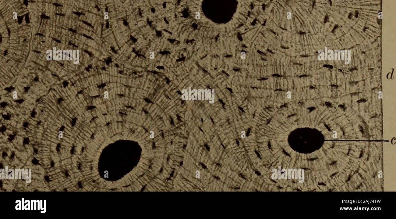
Two types of bone tissues in cross section of a long bone : Cross section of a bone : To the left is muscle tissue, and to the right is bone marrow. The development of a sliding mechanism for cutting machines began as early as 1798 as a method to section large tissue specimens. Bone and bones / pathology*. Fetal leg, cross section, h&e, 40x (bone marrow in tibia and fibula, developing blood cells, sinusoid 37829 x 41067, megakaryocyte 37861 x 39647, 38143 x 39087, 39555 x 36969, 31707 x 18214). At the outer regions of the section, you can see a dense, thick layer of compact bone. A typical long bone shows the gross anatomical characteristics of bone. A cross section of any bone will demonstrate these two types of bones. This slide contained a cross section of a very small bone, and you are looking at the entire thickness of the shaft of the bone. The osteocytes are arranged in concentric rings of bone matrix called lamellae (little plates), and their processes run in interconnecting canaliculi. Histology sauropod vertebra picture of the week these pictures of this page are about:long bone cross section. Nigel harness, a uk histology technician with 26 years of experience in sectioning bone, advocates manual sectioning for paraffin embedded specimens because of the need to use varying levels of.
Spongy bone, also known as cancellous bone or trabecular bone, looks like a sponge under the microscope. Compact bone cross section courtesy: The arrows point toward the tumor. Nigel harness, a uk histology technician with 26 years of experience in sectioning bone, advocates manual sectioning for paraffin embedded specimens because of the need to use varying levels of. A typical long bone shows the gross anatomical characteristics of bone.
A Text Book Of Dental Histology And Embryology Including Laboratory Directions V4vr I V Lt Gt Lt V Y 1 F 4 F U V from c8.alamy.com Eventually, chevalier introduced the name microtome for these devices in 1839. Spongy bone is the osseous tissue, which fills the interior cavity of bones, consisting of mineralized bars called there are two ways to study bone histology. In addition to discussing the cellular constituents of bone and the architectural arrangement of their. The development of a sliding mechanism for cutting machines began as early as 1798 as a method to section large tissue specimens. If you are looking for the online quiz that this printable worksheet is based on, visit bone histology bone cross section. The cross section of a rectangular pyramid is a rectangle. Spongy bone, also known as cancellous bone or trabecular bone, looks like a sponge under the microscope. Decalcified compact bone looks completely different than compact bone that still has calcium salts in its matrix.
The wider section at each end of the bone is called the epiphysis (plural = epiphyses), which is filled with spongy bone.
Histology sauropod vertebra picture of the week these pictures of this page are about:long bone cross section. This slide contained a cross section of a very small bone, and you are looking at the entire thickness of the shaft of the bone. A tissue cut in the long direction is called a longitudinal section (l.s.), and one cut perpendicular to this is a cross section (c.s. Spongy bone is the osseous tissue, which fills the interior cavity of bones, consisting of mineralized bars called there are two ways to study bone histology. To the left is muscle tissue, and to the right is bone marrow. Or x.s.), or transverse section (t.s.). Slides have to be made this way because the matrix of bone is too hard to be cut with a knife as the other tissues are. There are two ways to study bone histology. This is a cross section through decalcified bone. At the outer regions of the section, you can see a dense, thick layer of compact bone. Most features of bone (but not the canaliculi, If you are looking for the online quiz that this printable worksheet is based on, visit bone histology bone cross section. The arrows point toward the tumor.
At the outer regions of the section, you can see a dense, thick layer of compact bone. Decalcified compact bone looks completely different than compact bone that still has calcium salts in its matrix. There are two ways to study bone histology. While it is not as hard as compact bone, spongy bone plays an important role of protecting the marrow where blood cells are produced. Histology of the haversian system (osteons, lamellae, canaliculi, volkmann's canals, and circumferential lamellae) in a ground bone section bone cross section.
Chapter 6 Page 5 Histologyolm 4 0 from stevegallik.org Later discussions in this chapter will show that bone is also dynamic in that its shape adjusts to accommodate stresses. There are a number of options available when the histologist is required to produce sections from bone or other calcified specimens. Cross and longitudinal sections (unstained). In the last decade, considerable technological improvements have been made to repair damaged bones and tissue, such as bone cross sections with implants for microscopic examinations. The central haversian canal, and horizontal canals (perforating/ volkmann's) canals contain blood vessels and nerves from the periosteum. Nigel harness, a uk histology technician with 26 years of experience in sectioning bone, advocates manual sectioning for paraffin embedded specimens because of the need to use varying levels of. Histology of the haversian system (osteons, osteocytes, canaliculi, lamellae, and cement line) in a section of demineralized bone. 'compact or cortical bone is usually thick dense bone that forms the outer shell cross sections of the bone when studied under the microscope reveal quite a different picture.at the outer regions of the section, you can see a dense, thick layer of compact bone.
Cross section of a bone :
Compact bone cross section courtesy: While it is not as hard as compact bone, spongy bone plays an important role of protecting the marrow where blood cells are produced. The slide labelled rabbit femur displays decalcified bone from which the mineral has been removed, leaving behind cells and organic matrix. Cross and longitudinal sections (unstained). If you are looking for the online quiz that this printable worksheet is based on, visit bone histology bone cross section. Histology of the haversian system (osteons, lamellae, canaliculi, volkmann's canals, and circumferential lamellae) in a ground bone section bone cross section. Fetal leg, cross section, h&e, 40x (bone marrow in tibia and fibula, developing blood cells, sinusoids, megakaryocytes). Histology slide courtesy of william l. This is a cross section through decalcified bone. The inner portion of the bone is composed of trabecular bone and the intervening bone marrow. Spongy bone, also known as cancellous bone or trabecular bone, looks like a sponge under the microscope. Fetal leg, cross section, h&e, 40x (bone marrow in tibia and fibula, developing blood cells, sinusoid 37829 x 41067, megakaryocyte 37861 x 39647, 38143 x 39087, 39555 x 36969, 31707 x 18214). To the left is muscle tissue, and to the right is bone marrow.
Red marrow fills the spaces in the spongy bone bone cross section. In choosing a technique and processing method, consideration must be given to the type of investigation being carried out.